What Is a Normal Respiratory Rate? Adult Breathing Rate Explained
A resting adult respiratory rate is commonly around 12–20 breaths per minute, but age, activity, illness, pain and anxiety can change it.
Respiratory rate is one of the most useful vital signs because it often changes early in illness. It should be counted carefully rather than estimated from a monitor alone.
How to count respiratory rate
Observe chest or abdominal movement while the person is resting. One rise and fall counts as one breath. Counting for a full minute is useful when breathing is irregular.
What can make breathing faster?
Exercise, fever, pain, anxiety, asthma, pneumonia, sepsis, pulmonary embolism and metabolic problems can all increase respiratory rate.
What can make breathing slower?
Sleep, medications, neurological conditions and respiratory depression can reduce breathing rate. Slow breathing with reduced consciousness is particularly concerning.
Why respiratory rate can matter more than SpO₂
A patient may have increasing work of breathing or respiratory rate before oxygen saturation falls. That is why clinicians combine rate, effort, speech, mental status and SpO₂.
Look for work of breathing
Accessory muscle use, inability to speak full sentences, grunting, paradoxical movement and exhaustion can indicate significant respiratory distress regardless of the exact rate.
When should you seek medical help?
Seek urgent help for severe breathlessness, inability to speak normally, blue or grey skin, confusion, collapse or rapidly worsening breathing.
Why respiratory rate deserves more attention
Respiratory rate is one of the most informative vital signs and one of the most commonly estimated rather than measured. A rising rate can be an early sign of physiological stress before oxygen saturation or blood pressure becomes abnormal.
Breathing rate should therefore be counted and interpreted alongside depth, effort, rhythm, speech, oxygen saturation and mental status.
What is a typical adult resting rate?
Many clinical references describe roughly 12–20 breaths per minute as a common resting adult range. This is a population reference, not a diagnosis. Fitness, sleep, pregnancy, fever, pain, medication and illness can shift the rate.
The key is whether the rate fits the context and whether it is changing.
How to count respiratory rate accurately
Count chest or abdominal rises for a sufficient period, ideally without drawing attention to the measurement because conscious awareness can change breathing. Irregular patterns may require a longer count.
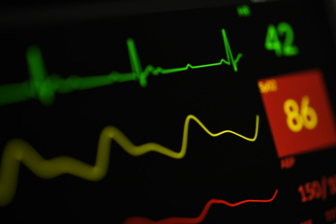
Do not rely only on the monitor. Automated respiratory-rate estimates can be inaccurate depending on the technology and movement.
Tachypnea: fast breathing
Fast breathing can occur with fever, pain, anxiety, exercise, asthma, pneumonia, pulmonary embolism, sepsis, metabolic acidosis, shock and many other conditions. The rate alone does not identify the cause.
Severity depends on work of breathing, oxygenation, fatigue, mental status and the underlying condition.
Bradypnea: slow breathing
Slow breathing can occur during sleep or in trained athletes, but can also indicate opioid or sedative effect, neurological injury, severe fatigue or other problems. A slow rate with reduced consciousness is especially concerning.
Respiratory rate versus oxygen saturation
SpO₂ and respiratory rate measure different things. Saturation can remain normal while the patient works very hard to maintain it. Conversely, a low saturation may occur with relatively normal respiratory rate.
Never let one normal value cancel out obvious respiratory distress.
Work of breathing matters
Accessory muscle use, inability to speak full sentences, nasal flaring, retractions, tripod positioning and visible exhaustion can indicate severity even if the numerical rate is only mildly abnormal.
Patterns and rhythm
Breathing can be regular, irregular, shallow, deep or periodic. Abnormal patterns may reflect neurological, metabolic or toxicological problems. The pattern can be as important as the count.
Respiratory rate in infection and sepsis
Tachypnea can be an early marker of serious infection or metabolic stress. A patient with fever, confusion and rising respiratory rate deserves careful assessment even if oxygen saturation is normal.
Respiratory rate in metabolic emergencies
Deep rapid breathing can occur when the body compensates for metabolic acidosis, such as diabetic ketoacidosis. Treating it as “anxiety” without considering metabolic causes can delay care.
Respiratory rate in children
Normal ranges vary substantially with age, so adult thresholds should not be applied to children. In pediatric assessment, work of breathing, behavior and perfusion are especially important.
Respiratory rate in pregnancy
Pregnancy changes respiratory physiology, but marked breathlessness or significant tachypnea should not simply be attributed to pregnancy. Chest pain, syncope, unilateral leg swelling or severe symptoms require prompt assessment.
Field scenario: normal SpO₂, abnormal breathing
A patient has SpO₂ 97% but breathes 32 times per minute with increasing fatigue. The normal saturation does not make the patient low risk. The respiratory rate and work of breathing show significant physiological stress.
Field scenario: anxiety versus serious illness
A person with panic symptoms is tachypneic, but so is a patient with pulmonary embolism or sepsis. Anxiety should be considered only after appropriate assessment of red flags, history and physiology.
Common mistakes
- estimating instead of counting
- ignoring depth and work of breathing
- assuming normal SpO₂ means breathing is normal
- using adult ranges for children
- not repeating the rate when the patient changes
Key points
- Count respiratory rate rather than estimating it.
- Interpret rate with effort, depth, rhythm and oxygenation.
- A normal oxygen saturation does not exclude respiratory failure.
- Age, pregnancy, fever, pain and medications affect breathing rate.
- Trend is often more useful than one isolated count.
FAQ
Is 22 breaths per minute always dangerous? No, but it is above many adult reference ranges and should be interpreted with the clinical picture.
Can anxiety increase respiratory rate? Yes.
Can breathing be dangerous even with normal SpO₂? Yes. Work of breathing and ventilation can deteriorate before oxygen saturation changes.
Related ActuMedic guides
How respiratory rate fits into a complete breathing assessment
Respiratory rate becomes much more useful when it is combined with the rest of the breathing assessment. Clinicians look at chest movement, depth, rhythm, air entry, breath sounds when appropriate, ability to speak, skin color, mental status and the patient’s own description of breathlessness. A person breathing 24 times per minute while speaking normally and improving after rest presents a different picture from someone at the same rate who is using accessory muscles, becoming drowsy and unable to finish a sentence.
For EMS clinicians, the practical question is not simply whether a rate falls inside or outside a reference range. The question is whether ventilation appears effective, whether the work required to maintain it is sustainable, and whether the pattern is improving or deteriorating over repeated assessments.
Why reassessment is essential
A single respiratory rate is a snapshot. Repeating the measurement after positioning, oxygen or other locally authorized treatment can reveal whether the patient is responding. A rate falling from 34 to 24 with easier speech and reduced accessory-muscle use may indicate improvement; a rate falling from 34 to 10 while consciousness deteriorates may indicate fatigue or respiratory depression rather than recovery.
Document the time and context of repeated observations. Trend interpretation is especially important during transport because a patient’s appearance can change before a monitor generates an alarm.
Respiratory rate and ventilation are not the same thing
Minute ventilation depends on both respiratory rate and the volume of air moved with each breath. A patient can breathe rapidly but very shallowly and still ventilate poorly. Another patient can have a relatively normal rate but inadequate tidal volume because of fatigue, neuromuscular weakness, medication effect or chest injury.
This is one reason rate should never be used as a substitute for observing depth and effectiveness. Where capnography is available and within local practice, it may add information about ventilation, but it also requires clinical interpretation and does not replace direct assessment.
What respiratory rate can tell you about circulation
Tachypnea is not limited to lung disease. Shock, blood loss and poor perfusion can drive faster breathing as the body responds to metabolic stress. In a patient with trauma, gastrointestinal bleeding or suspected sepsis, an increasing respiratory rate may therefore be an early deterioration signal even when the lungs are not the primary problem.
Look for accompanying changes such as tachycardia, altered mental status, cool or mottled skin, delayed capillary refill where appropriate, falling blood pressure or increasing weakness. No single sign proves shock, but a cluster of worsening findings deserves escalation.
Respiratory rate and pain
Pain commonly increases breathing rate, particularly after trauma, abdominal pain or pleuritic chest pain. The pattern may be shallow because deeper breaths hurt. Do not automatically attribute tachypnea to pain, however, until serious respiratory, circulatory and metabolic causes have been considered.
Reassessment after appropriate pain management can be informative. Improvement in rate and depth may support pain as a contributor, while persistent or worsening abnormalities should prompt renewed assessment.
Respiratory rate and fever
Fever can increase respiratory rate through higher metabolic demand. In infection, however, marked or rising tachypnea can also signal systemic illness. The combination of fever, confusion, hypotension, reduced urine output or rapidly worsening breathing deserves urgent clinical attention.
Do not use a normal oxygen saturation to dismiss a rising respiratory rate in a patient who otherwise appears unwell.
Special considerations at altitude
At altitude, lower oxygen pressure can increase breathing rate and reduce oxygen saturation compared with sea-level values. Visitors who are not acclimatized may notice faster breathing with exertion. Severe headache, confusion, marked breathlessness at rest or neurological symptoms are not normal acclimatization and require prompt assessment.
Opioids, sedatives and respiratory depression
Opioids and other sedating substances can reduce respiratory drive. A slow rate accompanied by reduced consciousness, shallow breathing, cyanosis or poor air movement is a high-risk pattern. Counting rate alone can underestimate severity if breaths are ineffective.
EMS assessment should follow local toxicology and airway protocols, with close attention to airway patency, ventilation, oxygenation and response to any authorized intervention.
Neurological causes of abnormal breathing
Brain injury, stroke, seizure, raised intracranial pressure and other neurological conditions can alter respiratory rate or pattern. Irregular breathing with reduced consciousness is particularly concerning. In these situations, airway protection, ventilation and neurological reassessment are linked priorities.
What to communicate in an EMS handover
A useful respiratory handover includes more than the latest number. State the initial rate, subsequent trend, work of breathing, oxygen saturation, relevant breath sounds if assessed, oxygen or ventilation support provided, and the patient’s response. Mention whether the patient can speak normally and whether mental status changed.
For example, “Respiratory rate increased from 24 to 32, now speaking in short phrases with increasing accessory-muscle use despite unchanged SpO₂” communicates deterioration more clearly than “RR 32.”
A practical reassessment checklist
- Repeat the respiratory rate rather than copying the previous value.
- Reassess depth, rhythm and work of breathing.
- Check ability to speak and changes in mental status.
- Review oxygen saturation in context and verify signal quality.
- Look for circulatory or metabolic clues that may explain tachypnea.
- Record the response to positioning or locally authorized treatment.
- Escalate when the overall trajectory is worsening.
More questions about respiratory rate
Should respiratory rate always be counted for 60 seconds? A full minute is particularly useful when breathing is irregular, very slow or difficult to count. Shorter validated counting methods may be used locally for regular breathing, but accuracy matters more than speed.
Can a person have respiratory failure with a normal rate? Yes. Depth, fatigue, gas exchange, mental status and the underlying condition matter. A normal numerical rate does not guarantee effective ventilation.
Why can respiratory rate change before blood pressure? Breathing responds rapidly to metabolic demand, acidosis, pain, fever and physiological stress, so it can become abnormal before later signs of decompensation appear.
Should a smartwatch respiratory rate be treated as a clinical measurement? Consumer devices can show trends but are not a substitute for clinical assessment. Unexpected or concerning readings should be confirmed and interpreted with symptoms.
Documentation: make the respiratory trend visible
Good documentation allows another clinician to reconstruct what happened. Record the measured rate rather than terms such as “breathing fast” alone, and add clinically meaningful descriptors such as shallow, irregular, labored or unlabored when observed. If the patient receives oxygen, ventilation support, bronchodilator therapy or another locally authorized intervention, record the timing and subsequent reassessment.
A sequence such as “RR 26 at first contact, 31 before departure, 34 during transport with increasing accessory-muscle use” immediately communicates trajectory. This is more useful for continuity of care, clinical review and education than repeating the same copied vital-sign set.
How to avoid anchoring on a respiratory diagnosis
Fast breathing often makes clinicians think first about asthma, pneumonia or anxiety. Those are possibilities, but tachypnea can also accompany pulmonary embolism, shock, metabolic acidosis, sepsis, pain, fever and other conditions. A structured assessment reduces the risk of stopping at the first plausible explanation.
Ask whether the history, examination and vital-sign pattern all support the working impression. If they do not, reopen the differential and escalate according to local practice.
Bottom line for EMS learners
Respiratory rate is simple to obtain but clinically rich when measured accurately. Learn to notice the number, the pattern, the work required to breathe and the direction of change. Avoid treating 12–20 as a pass/fail test. The patient in front of you, their baseline and their trajectory determine the significance.
For students and working clinicians, one of the most useful habits is to measure respiratory rate early and repeat it deliberately. That habit creates a baseline, improves handover and can expose deterioration that might otherwise be missed.
Respiratory rate: a final practical example
Consider two adults breathing 26 times per minute. The first has just walked quickly, is comfortable, speaking normally and settles to 18 after rest. The second is sitting still with fever, new confusion and increasing work of breathing. The numerical rate is the same at the first measurement, but the second patient has a much more concerning clinical pattern.
Now consider a patient whose rate falls from 30 to 12. That may look like improvement, but if the patient is becoming drowsy and breaths are shallow, the change may instead reflect fatigue or respiratory depression. Reassessment must therefore include depth, effort, mental status and oxygenation, not simply whether the rate moved closer to a reference range.
For EMS learners, respiratory rate is an excellent exercise in clinical reasoning because it forces you to connect a simple observation with physiology. Measure it accurately, repeat it when the patient changes and communicate the trend. Severe breathlessness, altered consciousness, cyanosis, exhaustion or rapid deterioration requires urgent escalation according to local emergency protocols.
Sources
General health information only. Measurements should be interpreted alongside symptoms, baseline health and professional assessment.